Tuesday 16 February 2016

Modern vs. Traditional; The exact way to Determine Corneal Diameter in Cataract surgery

Corneal diameter
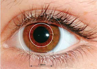
Corneal diameter is determined by the horizontal White to White (WTW), as it has significant clinical implications for the successful completion of the cataract surgery.  The mean horizontal white-to-white (WTW) corneal diameter is needed to determine the diameter of the intraocular lenses to be implanted after extracting the affected clear lenses in the cataract surgery. Traditionally, calipers are used to measure the diameter of the cornea manually. With mechanization and automation in every field, IOLMasters have taken up this task.The present study tries to compare whether the traditional means of manual measurement, using calipers is effective or the IOLMasters are fruitful in serving the task?


microphthalmia
Measurements related to microphthalmia, relative anterior microphthalmia, microcornea, macrophthalmia, and macrocornea are crucial to determine the corneal diameter as surgeons rely on the measured corneal diameter for sizing the intraocular lenses. However, no two sources of information provide exact estimation of the normal horizontal corneal diameter, as most of these texts are written when the automated means of IOLMasters were not available.  While some sources define microcornea in the range of 10.0 to 11.0 mm the other sources put it around 12.5 to 13.0 in adults. The current accepted standards of normal WTW at present is greater than 11.0 mm and less than 13.0 mm.

Authors Tina H Chen and Robert H Osher for the present study have made a retrospective chart review of cataract surgery for 516 patients by a senior surgeon at the Cincinnati Eye Institute (CEI) from September 2010 to November 2012. The patients had cataract surgeries in one or both eyes Pre-operative measurements with the IOLMaster, including unilateral or bilateral measurement of the horizontal WTW corneal diameter were recorded. Manual caliper measurements were performed by adjusting the calipers under the microscope by placing each tip and the data was analyzed using SPSS.

Corneal diameters measured with the calipers and the IOLMaster did not correlate significantly with age as assessed with both linear correlations. Males had slightly but significantly larger corneal diameters as compared to women with both calipers. There was no difference in corneal diameters of left versus right eye with both calipers (p=0.09) and IOLMaster

The study found that WTW measurements produced by the IOLMaster were slightly but significantly smaller than caliper WTW measurements, so these two devices should not be considered interchangeable when future population studies are conducted. The study concludes that macrocornea as a WTW measuring larger than 13.2 mm and supports the current definition of microcornea as WTW measuring 11.0 and smaller.
Friday 12 February 2016

The other side of the Lasik Surgery

laser eye surgery Laser in situ keratomileusis, popularly known as LASIK is a laser eye surgery used to correct vision refractions to correct myopia, hyperopia, and astigmatism. Under the ideal conditions, patients that undergo lasik surgery get well soon. Patients may develop post-operative complications if the surgery is not properly conducted.   The major among the complications would be development of Corneal epithelial cell growth on the visual axis that severely damages the vision. In such cases, patient’s cornea melts away gradually, leading to complete loss of vision if the epithelial growth is not properly handled.

Corneal flap is the first step in the Lasik surgery, which is created by placing a ring over the cornea. This allows some space between the flap and the stroma, leading to corneal epithelial cell growth. Corneal peripheral structure will be cut for this purpose. Lifting of the cornea experiences flap manipulation and cellular migration, leading to ‘corneal epithelial’ growth.
Corneal epithelial cell growth

Hassan Aboumerh et al in their study observed the case of a 50 year old woman that developed corneal epithelial growth 15 years after the initial surgery.  She was hurt in her garden when a twif poked her eye and she was admitted as an emergency case. Slit lamp examination of the cornea after the initial injection of conjunctiva showed corneal abrasion of the epithelium temporally surrounded by mild corneal edema. The flap was intact and its edges were not involved. Doctors administered tobramycin/dexmathesaone ophthalmic drops to the patient and advised her to be present after a week. Patient recovered quickly. Keeping the potential threat of the in growth to the optic nerve, the doctors have removed the epithelial growth. The patient was put on ofloxacin and prednisolone acetate drops and was observed for a day. She returned with a clear visual axis and a smooth flap. One month later, there was no return of epithelial ingrowth and the patient's vision was still 20/20 in both eyes.

Tuesday 2 February 2016

Ensuring Perfect Vision Care with Right Lens

Many varieties of lenses, including ophthalmic lenses, contact lenses, intraocular lenses, filter and low vision instruments are often used to treat eye related diseases, to correct the refraction errors and to attain an improved and better vision. Ophthalmic optical lenses are prescribed based on the basic laws of geometrical optics and visual optics issues. Light plays a crucial role in the eye care as the wave length of the light increases visual light transmittance and decreases aberrations, and allows the lenses to project a clearer vision. Properties of the lenses are tested depending upon their physical and chemical characteristics and UVR and light transmittance analyses of ophthalmic optics lens are crucial in terms of eye health. Lenses were tested for their transmittance properties depending on the physical, chemical characteristics. The UVR and light transmittance analyses of ophthalmic optics lens are crucial in terms of eye health.
Ophthalmic optical lenses

Physical characteristics involve the curvature of surfaces, vergence, categories of lens form, the diameter (R), base curve (BC), diopter (D), sagittal depth (S), surface-type (spherocylindrical), edge thickness (ET), optical center (OC), focal point(F), focal power (D) and refractive index (n) of the lenses. The chemical characteristics of ophthalmic lenses are classified as mineral lenses (crown, flint, borosilicate, heavy flint) and organic lenses (CR-39), high index, polycarbonate and trivex). Physical parameters such as diameter (55–70 mm), center thickness (1–5 mm), refractive index (1.50– refractive index (1.50–1.90), diopters (D), base curvature (BC) and focal point (F) of lenses are also important to determine the spectral transmittance results. According to the results, the UVR and visible-light transmittance within the spectrum depends not only on the composition of the eye-glasses but also on the lens thickness, the composition of the lens material, the index numbers and coating characteristics of the lenses.
UVR

The UVR and visible-light transmittance within the spectrum depends not only on the composition of the eye-glasses but also on the lens thickness, the composition of the lens material, the index numbers and coating characteristics of the lenses. The physical, chemical and optical characteristics should be evaluated prior to transmittance assessment of lenses. Since long ultraviolet radiation exposure causes various eye diseases, light transmittance value in the ultraviolet radiation region should be close 0%. Use of ophthalmic lenses with 85% and higher transmittance was suitable to ensure perfect eye care.


Attaining Perfect Vision with CCC technique in Cataract Surgery


Traditionally, the surgeons were opening the capsule of the lens of an eye to treat cataract and they used to extract the lens and the capsule to prevent any possible inflammation after surgery due to leftover parts.
Eye Cataract
Modern cataract surgeries are no longer extracting the entire capsule with the introduction of a modern technique called continuous curvilinear capsulorhexis (CCC). Modern day surgeons are able to remove the entire residual left after the surgery with this technique. Surgeons used to remove the affected layer along with the capsule in the past. With the introduction of Intraocular Lens (IOL), the ophthalmologist is able to remove the entire affected layer while retaining the capsule. The implanted lens however, may vary from surgeon to surgeon and depending upon the type of IOL implanted; there will be a 360◦ optic overlap. While excessive overlap may cause a postoperative hyperopic shift, incomplete overlap is associated with IOL tilt, causing lack of clarity in the vision. 

Intraocular lens(IOL)
According to a study called Impact of Capsulorhexis Morphology on the Predictability of Intraocular Lens Power Calculations, Jonathan M Davidorf argues that compared to acceptably shaped CCCs, a perfectly centered, perfectly round anterior capsulotomy will improve the predictability of the effective lens position. The study reviewed 175 random cataract surgery videos (acrylic IOL) to assess the impact of capsulorhexis (CCC) morphology on predictability of intraocular lens (IOL) calculations. The study found no significant difference in variance and concludes that there appears to be no relationship between CCC morphology and IOL calculation predictability.